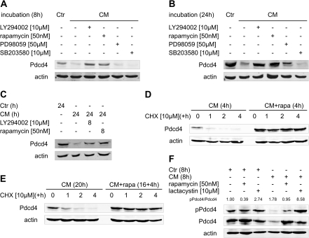
Fig. 4.
CM-induced Pdcd4 protein degradation requires intact PI3K–mTOR signaling. MCF7 cells were incubated with CM in combination with LY294002 [10 μM], rapamycin [50 nM], PD98059 [50 μM] or SB203580 [10 μM] for (A) 8 h or (B) 24 h. (C) MCF7 cells were incubated with CM or Ctr for 24 h. LY294002 [10 μM] or rapamycin [50 nM] were added for the last 8 h of the incubations. (D) MCF7 cells were incubated with CM (±rapamycin [50 nM]) for 4 h and (E) pre-incubated with CM for 16 h before incubations continued in the presence or absence of rapamycin [50 nM] for 4 h. Then, CHX [10 μM] was added to block translation and incubations continued for 1, 2 or 4 h (D + E). (F) MCF7 cells were incubated with Ctr or CM for 8 h in combination with rapamycin [50 nM] or lactacystin [10 μM]. Whole-cell extracts were subjected to western blot analysis and probed with the indicated antibodies. Blots are representative of at least three independent experiments. Ratios of pPdcd4 to Pdcd4 were determined after densitometric analysis of the expression and are presented as means of four independent experiments (F).

