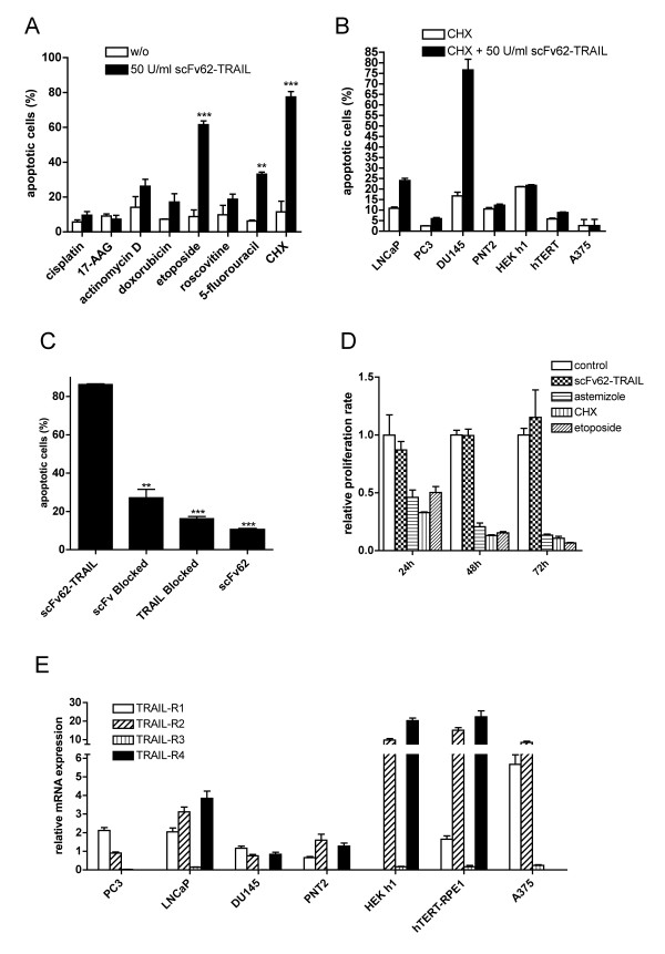
Figure 4.
KV10.1-specific apoptosis induction. A) Annexin/PI staining and flow cytometry analysis of DU145 cells treated with scFv62-TRAIL in combination with different chemotherapeutic agents: cisplatin (10 μM), 17-AAG (5 μM), actinomycin D (800 nM), doxorubicin (1.8 μM), etoposide (50 μM), roscovitine (10 μM), 5-fluorouracil (100 μM), CHX (5 μg/ml) (n = 2). B) Cell lines were treated for 18 h with 50 U/ml scFv62-TRAIL in combination with 5 μg/ml CHX and analyzed for apoptosis with Annexin/PI staining and flow cytometry (n = 2). C) Blocking assay: DU145 were treated with scFv62-TRAIL, scFv62-TRAIL pre-incubated with anti-TRAIL antibody or scFv62-TRAIL pre-incubated with antigen, in all cases in the presence of CHX. Alternatively, cells were pre-incubated with scFv62 for 1 h and then treated with scFv62-TRAIL and CHX. As a control, DU145 cells were treated with scFv62 preparation. Apoptosis induction was analyzed with Annexin/PI staining and flow cytometry (n = 3). D) Proliferation assay: DU145 cells were treated with 50 U/ml scFv62-TRAIL, astemizole (4 μM), CHX (5 μg/ml) or etoposide (5 μM) and proliferation was measured after 24, 48 and 72 h (n = 3). E) Quantitative real-time PCR analysis of the four TRAIL receptors.

