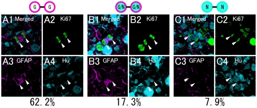
Figure 1. Characterization of dividing cells in the neonatal dentate gyrus.
P5 mice were fixed and then processed for immunohistochemistry. The prepared slices were stained with Ki67 (A2, B2, C2), GFAP (A3, B3, C3) and Hu (A4, B4, C4). A: A Ki67+ dividing cell pair expresses GFAP but not Hu. B: A Ki67+ dividing cell pair expresses both GFAP and Hu. C: A Ki67+ dividing cell pair expresses Hu but not GFAP. Scale bar, 10 µm.

