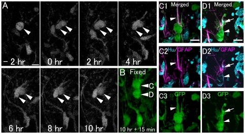
Figure 5. Asymmetric division of eGFP+ cells to produce a GFAP+ radial type cell and a neuronal cell.
A: Time-lapse imaging of GFP+ cell division in a hippocampal slice from a P6 GFAP-eGFP Tg mouse. Also see Video S3. B: 10 hours after cell division, the slice was fixed and then processed for immunohistochemistry. Optical images (C, D): The cells in B indicated by arrowheads C and D are located at different levels of the Z-axis. Each cell is shown separately in different single optical images of C and D. The GFP-positive cells (arrowheads C and D) in B correspond to those indicated by arrowheads in C and D, respectively. One GFP+ daughter cell has a radial process and an astrocytic cell marker (GFAP+). Another GFP+ daughter cell expresses GFAP and the neuronal marker Hu, suggesting a neuronal lineage-commitment. Scale bar, 10 µm.

