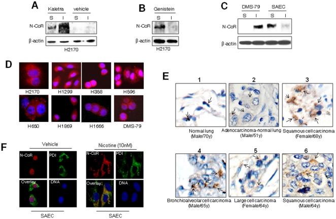
Figure 2. Loss of N-CoR in NSCLC cells is linked to misfolding.
A, B & C, Solubility (S)/insolubility (I) of N-CoR or β-actin in Kaletra (A) or genistein (B) treated H2170 cells, as well as in DMS-79 or SAEC cells (C), was determined by protein solubility assay. D, Subcellular distribution of N-CoR (red signal) in NSCLC (H2170, H1299, H358, H596, H650, H1869 and H1666) and SCLC (DMS-79) cells was determined by immunoflorescence assay performed with N-CoR antibody. DNA (blue signal) was stained with DAPI. E, Subcellular distribution of N-CoR in histologically confirmed human primary NSCLC tissue sections (panel 2–6) and normal human lung tissue section (panel 1) was determined by immunohistochemical (IHC) assay performed with N-CoR antibody. The brownish staining in the cytosol pinpointed by black arrow represents the N-CoR signal (panels 2–6). N-CoR staining in normal lung tissue section is visible as black dots overlapping the nucleus (panel 1). The second panel demonstrates a cross section that contains both cancer tissue (left half) and normal lung tissue (right half) side by side. F, Subcellular distribution of N-CoR and PDI in SAEC cells treated with vehicle or nicotine for 48 hours was determined by confocal microscopy.

