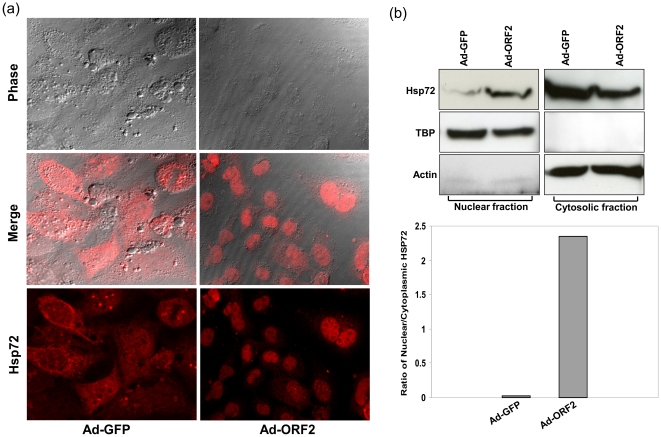
Figure 6. ORF2 increases nuclear accumulation of Hsp72.
(a) Huh7 cells grown on cover slips were infected with Ad-GFP or Ad-ORF2, fixed and stained with anti-Hsp72 antibody at 72 hpt. Cells were imaged by confocal microscopy and composite images were created using IMAGE J software. (b) Western blot of Hsp72 protein expression in cytosolic and nuclear fractions of Ad-GFP and Ad-ORF2 infected Huh7 cells at 72 after transduction. Cytosolic actin and nuclear TBP were used for equal loading. Signal intensities of Hsp72 for both, nuclei and cytoplasm were quantified and normalized by appropriate loading controls. The nuclear/cytoplasmic ratio was calculated as described in methods.

