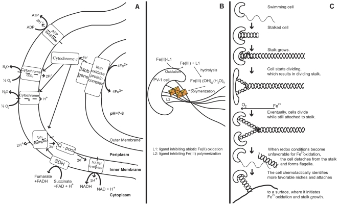
Figure 4. Conceptual iron oxidation model in relation to the life cycle in M. ferrooxydans PV-1.
A: Proteins potentially involved in the energy acquisition via Fe(II) oxidation through the outer and inner membrane as predicted from genomic analysis. The “Mob gene”, possibly located in the periplasm, represents the experimentally identified molybdopterin oxidoreductase Fe4-S4 region (SPV1_03948), which was extracted under iron oxidizing conditions as mentioned earlier. Its function may include the shuttling of electrons between outer and inner membrane. B: Biologically formed iron oxides are stored in the stalk of PV-1 as edge-sharing Fe-O6 octahedral linkages as previously described in [76]. As the cell performs Fe(II) oxidation, it rotates, which results in a twisted, coiled stalk. C: Schematic of the life cycle in PV-1. The cell moves in the environment until it identifies conditions suitable for Fe(II) oxidation. The flagella are discarded and stalk growth initiated. As the cell divides, the stalk becomes bifurcated, and each cell continues to form a stalk that is initially half the width as observed by [12]. When O2 concentrations exceed the maximum tolerable by PV-1, the cell detaches from the stalk and forms flagella to move to a better-suited niche, where the life cycle starts over.

