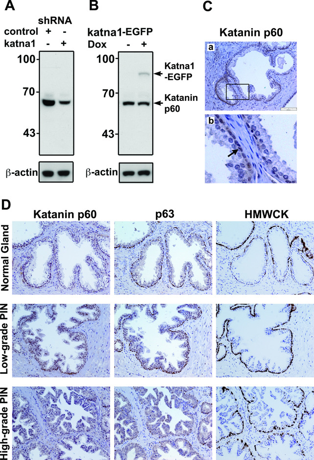
Figure 2.
Validation of katanin p60 antibody and immunohistochemical staining of normal prostatic glands and prostatic intraepithelial neoplasia. A. Western blot with katanin p60 antibody showed a 60 kDa band that was reduced after knockdown by KATNA1-shRNA but not by control shRNA in 293T cells. B. Katanin p60 antibody recognized the 85 kDa katanin p60-EGFP fusion protein. Expression of katanin p60-EGFP was achieved by transfection with a pTRE-KATNA1-EGFP in a Tet-On 293T cell line and induction with 1 µg/ml Doxycycline (Dox) for 24 hours. C. Immunostaining of normal prostate tissues with katanin p60 antibody showed that katanin p60 protein was weakly detectable in the epithelial cells. A boxed region is enlarged in panel (b). Note that arrow points to katanin p60 staining in the basal cell layer. D. Immunostaining of katanin p60 and basal cell markers (i.e. p63 and HMWCK) in normal prostatic glands, low-grade and high-grade PIN.

