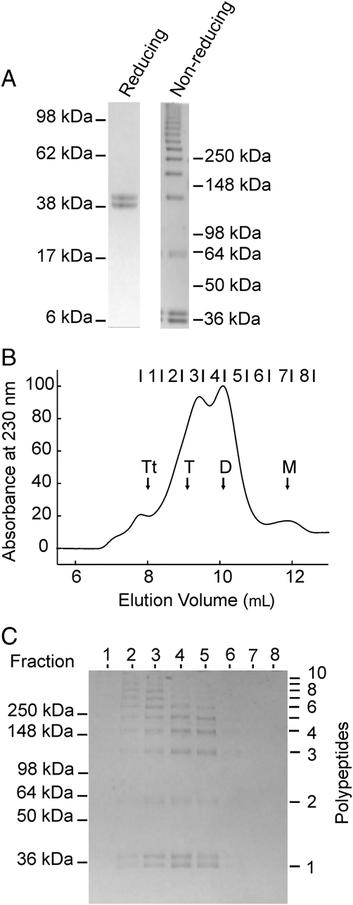
Figure 2.

Oligomeric structure of rat ficolin-B. (A) Purified ficolin-B separated by SDS-PAGE (4–12% gradient gel) under reducing and non-reducing conditions. Proteins were stained using Coomassie blue. (B) Gel filtration of ficolin-B on a Superdex-200 column. The elution positions of monomers (M), dimers (D), trimers (T) and tetramers (Tt) of rat ficolin-B are indicated. (C) Fractions collected across the gel filtration peaks were separated by gel electrophoresis on a 4–12% gradient SDS-polyacrylamide gel under non-reducing conditions, and proteins were stained with Coomassie blue.
