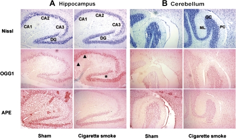
FIG. 3.
Nissl staining and immunohistochemical detection of OGG1 and APE1 in the hippocampus (A) and cerebellum (B) of a sham-exposed mouse and a CS-exposed mouse. CA1, CA2 and CA3 indicate the three hippocampal pyramidal cell layers and DG indicates the dentate gyrus. Note the heavy staining of OGG1 in the CA1 (arrowheads) and DG (star) regions of the hippocampus. GC, granule cell; ML, molecular layer; PC, Purkinje cell.

