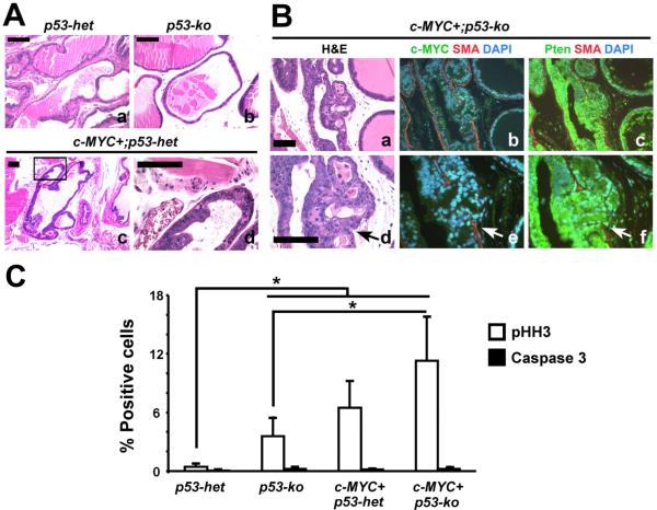
Figure 3. Modest cooperation between focal c-MYC expression and p53 haploinsufficiency or deficiency in the mouse prostates.
(A and B) H&E images showing the pathology in mutant mouse prostates. Note focal high grade PIN in c-MYC+;p53-het mouse prostates (`c' and `d' in A) and PIN with microinvasion (arrows) in c-MYC+;p53-ko prostate (`a' and `d' in B). Note breach in SMA, smooth muscle actin indicative of microinvasion in `b' and `e'. Panels `c' and `f' in B show Pten expression by immunofluorescence. Scale bars: 100μm. (C) Cellular proliferation and apoptosis in the mouse prostates are quantitated following immunohistochemistry for phospho-histone H3 (pHH3) and activated Caspase 3, respectively. N=3–5 per group. *P < 0.05.

