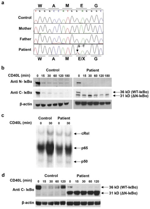
FIGURE 2.
Molecular analysis of the IKBA gene, and its effect on NF-κB signailing and IκBα protein expression. a: Chromatogram of the IKBA gene showing the heterozygous c.40G>T (p.Glu14X) mutation in the patient. Protein sequence for the healthy and mutated allele are represented above and below the DNA sequence respectively. b: Western blot analysis shows aberrant expression of an N-truncated IκBα protein in EBV-transformed patient B cells when a C-terminal specific antibody is used. The degradation of the aberrant N-truncated IκBα protein in response to CD40L stimulation is impaired. c: Impaired RelA and cRel binding in patient cells following CD40 stimulation. EBV transformed B cells were isolated from the patients and a normal control. Cells were stimulated in the presence of CD40L for 30 min. Cellular extracts were prepared and analyzed on the same gel by electrophoretic mobility shift assay for NF-κB binding activity. For supershift, all samples were incubated with anti c-Rel antibody prior to incubation with the labeled probe. One representative experiment of two is shown. d: In comparison to the control, resynthesis of wild-type IκBα protein level was notably reduced in the EBV-transformed B cell lines following stimulation with CD40L and in the absence of CHX. Cytoplasmic extracts were prepared from cells from a normal control and the patients and subjected to immunoblotting. Blots were probed with anti C-terminal IκBα antibody.

