Figure 2.

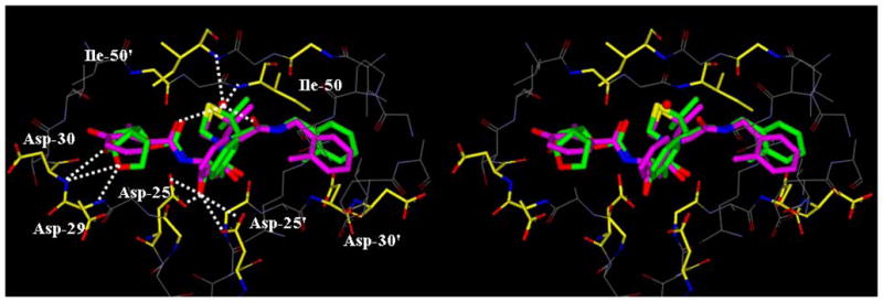
Structure of inhibitor 3b, modeled into the active site of HIV-1 protease, superimposed on the X-ray crystal structure of KNI-764. Inhibitor 3b carbons are shown in green and KNI-764 carbons are shown in magenta.

Structure of inhibitor 3b, modeled into the active site of HIV-1 protease, superimposed on the X-ray crystal structure of KNI-764. Inhibitor 3b carbons are shown in green and KNI-764 carbons are shown in magenta.