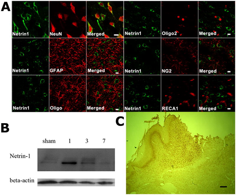
Figure 2. Early induction of endogenous netrin-1 in the neurons of the peri-infarct cortex following MCAO.
A, Confocal images showing double immunofluorescence staining of Netrin-1 (green) with cell type specific markers (red) including NeuN, GFAP, Oligo, Oligo2, NG2 or RECA at one day after MCAO. The results suggest that post-stroke endogenous netrin-1 was predominantly expressed by neurons. There was no netrin-1 immunoreactivity detected in GFAP-expressing astrocytes, oligodendrocytes, or RECA1-expressing blood vessels (n=3 per group). Scale bars: 10 μm. B, Western blots showing endogenous netrin-1 expression (rabbit-anti-mouse netrin-1) in sham, 1, 3, or 7 days after dMCAO. A robust induction of netrin-1 began as early as one day after MCAO, and lasted for one week. (n=3 per group). C, A low magnification view of endogenous netrin-1 expression suggests that it is mainly expressed in the peri-infarct cortex. Scale bar: 200 μm.

