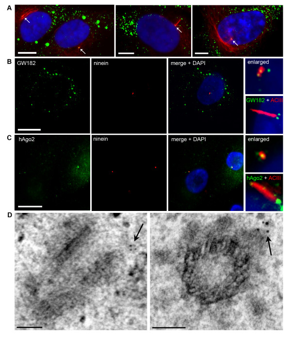
Figure 1.
RNA silencing proteins, GW182 and hAgo2, localize to the centrosome/basal body in human astrocytes throughout interphase. (A) Astrocytes double labeled with mouse anti-acetylated tubulin (red) and human GW182 antiserum 18033 (green) and counterstained with DAPI (blue). Left panel illustrates two daughter cells. Cells were seeded at a low density to allow the identification of daughter cells following the completion of mitosis (early G1). Each cell contains a centrosome with associated GW/P bodies (arrows). Centre panel illustrates a cell in G1/S phase (the period of primary cilium expression). One of the two GW/P bodies is demonstrated in this focal plane at the basal body (arrow). Right panel illustrates a late S/G2 cell with prominent chromatin condensation (DAPI) and a centrosome with two GW/P bodies (arrow). (B) GW182 and (C) hAgo2 localization to the centrosome and the basal body of primary cilia was examined by IIF using 18033 and mouse monoclonal 4F9 to recombinant hAgo2 [29] (green). Nuclei were marked by DAPI. The inset box in (B) shows the location of GW182 marked by mouse monoclonal 4B6 antibody and (C) hAgo2 mouse monoclonal antibody relative to the primary cilia. All IIF scale bars = 15 μm. (D) GW182 localizes to the pericentriolar region in immunoelectron micrographs. Arrows indicate the immunogold stained GW/P body foci. EM scale bars = 100 nm.

