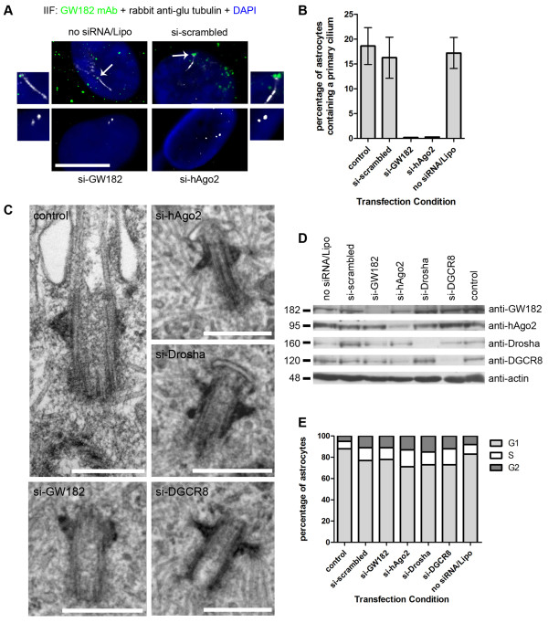
Figure 2.
SiRNA knockdown of RNAi components, GW182, hAgo2, Drosha and DGCR8, inhibits ciliogenesis in human astrocytes. (A) The primary cilium was examined by IIF using antibodies to glu tubulin (white) in astrocytes transfected with no siRNA/Lipofectamine, scrambled siRNA and si-GW182 and double stained for GW bodies using mouse monoclonal 4B6 antibody to GW182 (green). The primary cilium as marked by glu-tubulin (white) was also examined in astrocytes transfected with si-hAgo2 and double stained with mouse monoclonal 4F9 antibody to hAgo2 (green). Nuclei were stained by DAPI. The merge + DAPI images were enlarged to focus on the cilium and centrioles relative to GW182 and hAgo2 staining. Scale bar = 15 μm. (B) The percentage of astrocytes containing primary cilia were quantitated from 5 independent IIF experiments containing > 250 cells described in (A). Bars represent the standard deviation of the mean. (C) Ultrastructural analysis of control astrocytes, scale bar = 400 nm, si-GW182, si-hAgo2, si-Drosha and si-DGCR8 treated astrocytes scale bars = 500 nm. (D) Human astrocytes were transfected for 30 hours with 100 nM siRNA and Lipofectamine 2000 (Invitrogen), lysed and processed for western blot analysis. The cells in the no siRNA/Lipofectamine lane were treated with Lipofectamine 2000 only (no siRNA) and the control lane represented untreated cells. (E) Cell cycle analysis showing the percentage of human astrocyte cells in G1, S and G2 phase during normal growth conditions (control) and under 100 nM siRNA transfected conditions.

