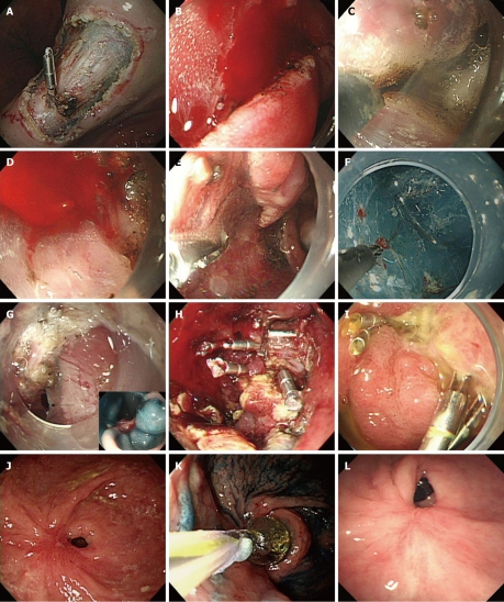
Figure 1.
Endoscopic views. A: Endoscopic view of exposed vessels on post endoscopic submucosal dissection (ESD) ulcer, showing hemoclipping for prevention of delayed bleeding; B, C: Endoscopic view of oozing of blood during ESD, showing immediate electrocoagulation by IT knife itself; D, E: Endoscopic view of pulsatile bleeding during ESD (D) and showing coagulation by hemostatic forcep (E); F: Endoscopic view shows that microvessels of the submucosal layer are cauterized by flex knife; G-I: Endoscopic view of jejunal loop side of G-Jstomy showing a perforation (arrow) seen during ESD for EGC of stoma of remnant stomach (G), and the view after closure of the perforation by endoclips (H). A follow-up endoscopy showed the healed perforation 2 wk after endoscopic closure (I); J-L: Endoscopic view shows severe antral stenosis 7 wk after gastric ESD (J). Endoscopic view of balloon dilation procedure (K). A follow-up endoscopic view showed relieved stenosis without any symptoms 3 mo after balloon dilatation (L). Ulcer induced stricture was detected at 4 wk after ESD.

