Figure 1.
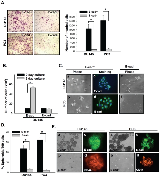
E-cad+ subpopulations isolated from DU145 and PC3 prostate cancer cells have a high invasive capacity and sustain stemness post-invasion. (A) Invasion was evaluated in E-cad+ and E-cad- subpopulations isolated from DU145 and PC3 cells. E-cad+ and E-cad-cells were plated in invasion chambers immediately after cell sorting. Twenty-four hours later, non-invaded (top-chamber) cells were removed, and the invaded cells were fixed and stained with crystal violet. *, P < 0.001. (B) Growth of invaded E-cad+ and E-cad- DU145 cells. After a 24 h invasion period, invaded E-cad+ and E-cad-cells were collected and plated under adherent culture conditions for 3 days and quantified. *, P < 0.001. (C) Phase-contrast photographs of invaded E-cad+ and E-cad-DU145 and PC3 cells after 3 days of culture, a, b, c and d, Invaded E-cad+ cells exhibited a holoclone-type morphology (10x) as well as E-cadherin expression (40x). e and f, phase-contrast photographs of invaded E-cad- cells after 3 days of culture. (D) In vitro quantification of prostate cell spheroids formed by invaded E-cad+ and E-cad- cells. The data are represented by the percentage of spheroids formed per 500 seeded cells ± SD. *, P < 0.001. (E) a and b, Phase-contrast images of DU145 (left panel) and PC3 (right panel) spheroids seeded by invaded E-cad+(top) and E-cad- (bottom) cells (10x). c and d, Images of spheroids immunostained with antibodies against E-cadherin (E-cad, green) and CD44 (red) (40x).

