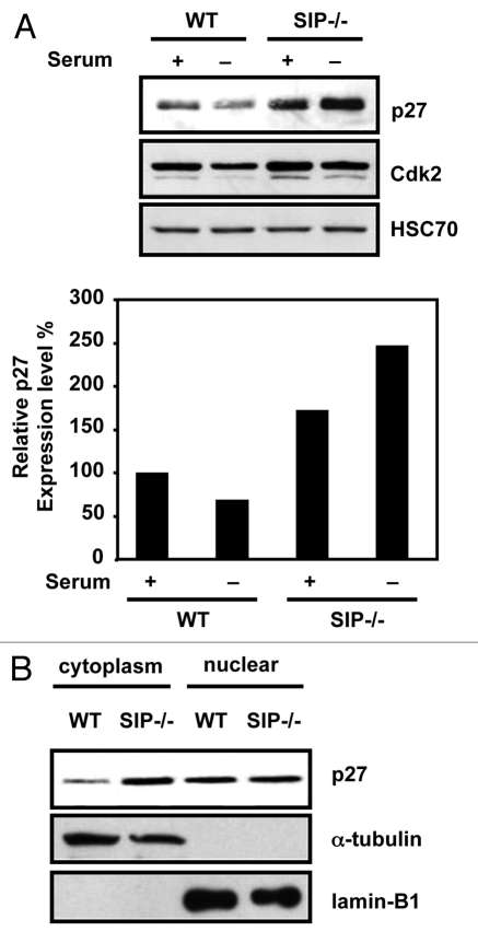
Figure 1.
Cytoplasmic p27 accumulates in SIp-deficient MeFs. (A) (top) Wild-type and SIP−/− MeFs were cultured in media containing 10% FCS (+) or 0.1% FCS (−) for 24 h. Cell lysates were analyzed by immunoblotting using antibodies specific for p27, Cdk2 or Hsc70. Representative experiment is shown. (Bottom) the degree of p27 expression was quantified by densitometric analysis with ImageJ software and expressed as a percentage of p27 expression in wild-type MEFs under normal growth conditions. Data are average of three time experiments. (B) Wild-type and SIP−/− MeFs cultured in complete media. Cell lysates were fractionated and analyzed by immunoblotting using antibodies specific for p27, Lamin B1 and α-tubulin, which acted as markers for the cytoplasmic and nuclear fractions, respectively.

