Figure 5.
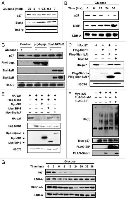
Siah1 is required for glucose limitation-induced p27 degradation. (A) Wild-type MEFs were cultured in media containing 10% dialyzed FCS and various concentrations of glucose. After 2 d, cell lysates were analyzed by immunoblotting using anti p27 or Siah1 antibodies. The membrane was reprobed with anti-HSC70 antibody as a control. (B) Wild-type MEFs cultured in complete media were released into glucose-free media. At the indicated times, lysates were analyzed by immunoblotting using antibodies for p27, Siah1 or LDH-A. (C) NIH3T3 cells were transiently transfected with plasmids encoding HA-Phyllopod or FLAG-Siah1ΔR and FLAG-Siah2ΔR as indicated. After 24 h, cell lysates were prepared from duplicate dishes for each transfection and analyzed by SDS-PAGE/immunoblotting using antibodies specific for p27, HA, FLAG and Hsc70 with ECL-based detection. (D) HEK293T cells were transiently transfected with plasmids encoding HA-p27, FLAG-Siah1 or FLAG-Siah1ΔR in various combinations as indicated (total DNA amount normalized). After 24 h, cell lysates were prepared from duplicate dishes of each transfection and analyzed by SDS-PAGE/immunoblotting using antibodies specific for HA and FLAG, with ECL-based detection. (E) HEK293T cells were transiently transfected with plasmids encoding HA-p27 (0.5 µg), FLAG-Siah1 (0.5 µg), Myc-SIP (0.5 µg), Myc-SIP-S (0.5 µg) or Myc-Skp2ΔF (0.5 µg) in various combinations as indicated (total DNA amount normalized). After 24 h, cell lysates were prepared from duplicate dishes of each transfection, normalized for total protein content (20 µg per lane) and analyzed by SDS-PAGE/immunoblotting using antibodies specific for HA-tag, FLAG or Myc, with ECL-based detection. (F) NIH3T3 cells were transiently transfected with plasmids encoding Myc-p27, FLAG-Siah1 and HA-SIP in various combinations as indicated (total DNA amount normalized). After 24 h, cells were treated with 1 µM MG132 for 6 h. Lysates were immunoprecipitated with anti-Myc antibody. Immunoprecipitates were divided into two partss and analyzed immunoblotting using anti-ubiquitin and anti-Myc antibodies. Expression of Myc-p27, HA-SIP in each total cell lysate was detected with anti-ubiquitin, -Myc, -HA and -FLAG antibodies. (G) Synchronized wild-type and Siah1a−/− MEFs were cultured in media containing 0.1 mM glucose and 10% dialyzed FCS. Cell lysates were prepared at the indicated times and subjected to immunoblotting with anti-p27 and LDH-A antibodies.

