Figure 6.
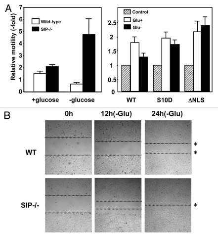
SIP regulates cell motility. (A) (Left) Serum-starved wild-type or SIP−/− MEFs were released into complete media or glucose-free media and allowed to migrate toward media containing 0.5% FBS or 10% FCS for 12 h. Colorimetric measurements were taken according to the manufacturer's instructions. (Right) 3T3 cells transfected with an empty pcDNA3 vector, pcDNA3 p27S10D or pc DNA3 p27ΔNLS were cultured in serum-starved media for 24 h, and were released into complete media or glucose-free media and allowed to migrate toward media containing 0.5% FBS or 10% FCS for 17 h. Colorimetric measurements were taken according to the manufacturer's instructions. (B) Serum-starved wild-type (WT) and SIP−/− MEFs were cultured in gelatin-coated 6-well plates to confluence and cells were introduced into media containing 2% FBS with (+Glu) or without glucose (−Glu). A wound area was mechanically induced by a single passage of a P200 tip across culture plate surface to confluent MEF monolayers. Cultures were continued for the indicated times and images obtained using light microscopy. Images were representative of six independent experiments. The dotted lines define the areas lacking cells.

