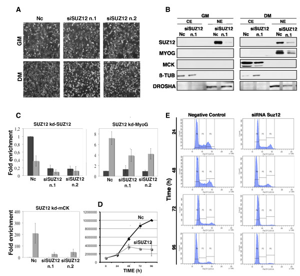
Figure 6.
Suz12 small interfering RNA (siRNA) impairs proliferation and differentiation in C2C12 cell lines. (A) Myoblasts were transfected with non-targeting siRNA (Nc = negative control) or siRNA against Suz12. The effect of siRNA on cell morphology was analysed in growth medium (GM) (48 h after transfection) and in differentiation medium (DM) (48 h after differentiation induction) by phase-contrast microscopy. (B) Immunoblot of Suz12, myogenin (MyoG) and muscle creatine kinase (mCK) was performed after Suz12 depletion (oligo no. 1) using the experimental design described in Figure 5A and the protein levels were analysed in cells cultured in GM and in DM (48 h after differentiation induction), respectively. Nuclear (NE) and cytosolic (CE) extracts were used for the analysis, with β-tubulin serving as a cytosolic and Drosha as a nuclear extract control. (C) The efficiency of Suz12 siRNA and the expression levels of MyoG and mCK were tested by real-time PCR in GM and in DM (48 h after differentiation induction) in Suz12-depleted cells. The transcription levels were normalised to Gapdh expression and represented as the average of three independent experiments ± SD. Fold enrichment was calculated in comparison to the negative control siRNA in GM. (D) Effect of Suz12 depletion on cell proliferation. The cells were counted 24 h, 48 h, 72 h and 96 h after siRNA transfection. Suz12 siRNA oligo no. 1 was used. Graph shows data from three independent experiments. Error bars represent the standard deviation (Nc = negative control). (E) Cell cycle profiles were analysed by fluorescence-activated cell sorting (FACS) after Suz12 siRNA delivery at the same time timepoints as in (D). P4 gate represents G1 phase, P5 represents S phase and P6 represents G2 phase.

