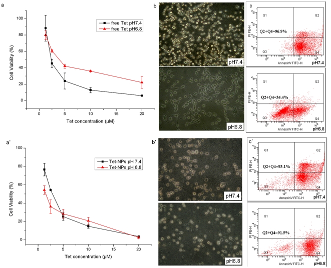
Figure 4. The PIPDR of MKN-28 cells to Tet ( Fig. 4.a–c ) and the reversion of PIPDR to Tet by incorporating it into PEG-PCL NPs ( Fig. 4.a′–c′ ).
The cytotoxicity of free Tet at different extracelluar pH values. pH 7.4 is the normal pH value used in cell culture while pH 6.8 is the pH value in most tumor tissues. Similar results can be found in other 3 different digestive cancer cells (Table.S4.). a. The morphological changes of MKN-28 cells at different extracelluar pH values. (400×). More cells remained normal morphology at lower extracellular pH. b. The apoptosis assay of MKN-28 cells treated with Tet at different extracelluar pH values. When the extracellular pH value decreased from 7.4 to 6.8, a prominent decrease in apoptotic ratio could be observed. a′ The cytotoxicity of Tet-NPs at different extracelluar pH values. pH 7.4 is the normal pH value used in cell culture while pH 6.8 is the pH value in most tumor tissues. Similar results can be found in other three different digestive cancer cells (Table.S4.). b′ The morphological changes of MKN-28 cells at different extracelluar pH values. (400×). The morphologic changes of MKN-28 cells were similar at both pH values. c′ The apoptosis assay of MKN-28 cells treated with Tet-NPs at different extracelluar pH values. The apoptotic ratios of the cells didn't change with the decrease of extracellular pH.

