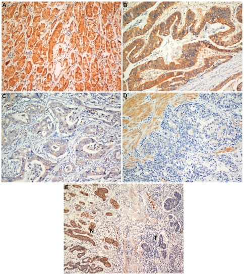
Figure 3. Immunohistochemical detection of the AP-2α protein expression in gastric cancer and surrounding non-tumor tissues.
(A) Normal gastric tissues, scored as AP-2α (+++); (B) Well-differentiated gastric cancer, scored as AP-2α (++) according to the criteria defined in material and methods section; (C) moderately differentiated gastric cancer, scored as AP-2α (+); (D) poorly differentiated gastric cancer, scored as AP-2α (−); (E) Immunostaining of gastric cancer and adjacent non-tumor tissues showing a sharp contrast between infiltrative tumor areas of negative staining and the adjacent tissue of positive staining. Original magnification: A–D×200; E×100.

