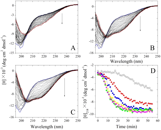
Figure 2. Far-UV CD spectra of Tau244–372 during fibril formation in the presence and absence of Pb2+ at 37°C.
10 µM Tau244–372 was incubated with 0–10 µM Pb2+ (A: 0 µM, B: 5 µM, and C: 10 µM). The arrows represented the incubation time increased gradually from 0 (the top, blue) to 54.4 min (the bottom, red). (D) Effect of Pb2+ on the relative change in the β-sheet content of Tau244–372 during fibril formation, studied by monitoring the CD signal at 218 nm. 10 µM Tau244–372 was incubated with 0–40 µM Pb2+ (black: 0 µM, red: 5 µM, blue: 10 µM, yellow: 20 µM, green: 30 µM, and magenta: 40 µM) in 30 mM phosphate buffer (pH 7.4) containing 1 mM DTT and 2.5 µM heparin.

