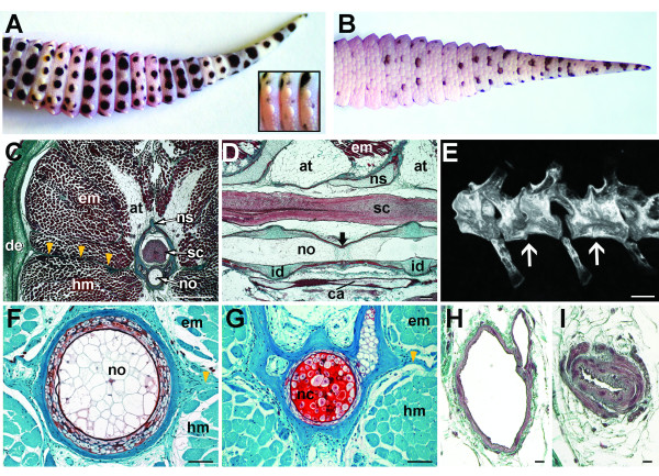
Figure 2.
Original anatomy of the gecko tail. Original tail anatomy. Eublepharis macularius. (A,B) In dorsal (A) and ventral views (B). Note the tubercles (inset) and the difference in pigmentation. (C-D, F-I) Serial sections (dorsal towards top of page). (C) Transverse section through distal third of the tail, stained with Masson's trichrome. The spinal cord (stained pink) is centrally positioned, enclosed within the neural canal of a vertebra (in this view stained green with red mottles). Surrounding the skeleton are bands of adipose tissue and skeletal muscle (stained red), enclosed by dermis (stained green) and epidermis (stained red). A longitudinal connective tissue septum radiates from the transverse process (yellow arrows). (D) Sagittal section through vertebral column, stained with Masson's trichrome. Ventral and parallel to the spinal cord is the notochord (in this view stained red with green mottles) and intervertebral disc (stained green). Ventral to the vertebral column is the caudal artery (stained red). The position of the intravertebral fracture plane indicated (black arrow). (E) Micro-computed tomography reconstruction of tail (caudal) vertebrae. Position of the intravertebral fracture planes indicated (white arrows). (F,G) Transverse sections through notochord in areas containing choroid tissue (F) and notochordal cartilage (G), stained with Safranin O. (H,I) Transverse sections through caudal artery before (H) and at the caudal arterial sphincter (I), stained with Masson's trichrome. at, adipose tissue; ca, caudal artery; de, dermis; em, epaxial musculature; hm, hypaxial musculature; id, intervertebral disc; nc, notochordal cartilage; no, notochord; ns, neural spine; sc; spinal cord. Scale bars: c, e = 500 μm; d = 100 μm; f = 50 μm; g = 20 μm; h,i = 10 μm; j-l = 20 μm.

