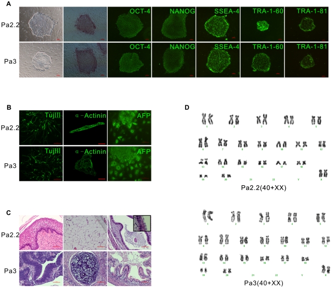
Figure 1. Isolation and characterization of two novel rhesus monkey parthenogenetic ESCs.
A, morphology and pluripotent markers. From left to right are Phase-contrast micrograph of an ESC colony growing on mEFs, Alkaline phosphatase staining, OCT-4, NANOG, SSEA-4, TRA-1-60 and TRA-1-81. Up is Pa2.2, down is Pa3; B, differentiation in vitro. From left to right are neuronal marker Tuj III, Cardiomyocytes marker α-cardiac actinin and Endoderm marker α-fetoprotein. Up is Pa2.2, down is Pa3; C, teratomas. Up is Pa2.2, from left to right are Squamous epithelium, adipose and respiratory epithelia (inset shows respiratory cilia). Down is Pa3, from left to right are neural tube, cartilage and intestinal epithelia; D, G-banding karyotypes. Up is Pa2.2, down is Pa3; Size bar (A, 100 µm; B and C, 50 µm).

