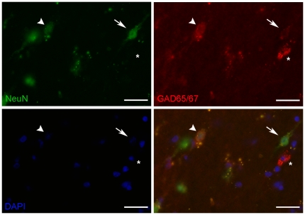
Figure 8. NeuN positive IWMNs express GABAergic markers.
Double-label immunofluorescence in adolescent (4.5 year old) rhesus macaque frontal pole coronal 14 µm sections shows co-localisation of NeuN (green) with GAD65/67 (red) in white matter neurons (DAPI staining for nuclei in blue). Some neurons show faint immunoreactivity for GAD65/67 (arrow) and some neurons express relatively more GAD65/67 and less NeuN (arrowhead) or no NeuN (asterisk). Scale bars = 25 µm.

