Figure 1.
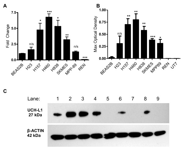
UCH-L1 expression is higher in NSCLC cell lines than in normal lung cells. A. Fold change of UCH-L1 mRNA in lung cancer cell lines compared to the normal lung cell line BEAS-2B (n = 3). B. Densitometry of the level of UCH-L1 protein detected by Western Blot relative to the level of β-actin detected (n = 3). C. Western Blot detection of UCH-L1 protein and β-actin loading control in different cell lines. Lanes as follows: 1 = H23, 2 = H157, 3 = H460, 4 = H838, 5 = BEAS-2B, 6 = MPP-89, 7 = REN, 8 = SKMES, 9 = UT-7.

