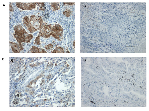
Figure 7.
UCH-L1 expression in adenocarcinoma and squamous cell carcinoma. A. Squamous cell carcinoma stained positive (i) and negative (ii) for UCH-L1. B. Adenocarcinoma positive (i) and negative (ii) for UCH-L1 expression. Brown staining indicates the presence UCH-L1. (Scale bar is equivalent to 25 μm).

