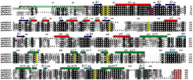
Figure 1. Multiple sequence alignment of POFUT1s.
Multiple sequence alignment of the GT65 family members CePOFUT1, HsPOFUT1, Gallus gallus POFUT1 (GgPOFUT1), Danio renio POFUT1 (DrPOFUT1) and DmPOFUT1. Secondary structure elements from the CePOFUT1 structure are shown, with α-helices in red and green for the N and C-terminal domains, respectively, and β-strands correspondingly in blue and cyan. Signal sequence of CePOFUT1 is highlighted in green while endoplasmic reticulum retention sequence for all POFUT1s are indicated in a pink box. Conserved cysteines forming disulphide bridges and N-glycosylated sites are shown in yellow and magenta, respectively. The boundaries of the N and C-terminal CePOFUT1 constructs are indicated in red for Thr26 and Ala382.

