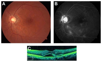
Figure 1.
(A) Before bevacizumab treatment, the color fundus photograph shows optic disc swelling, peripapillary hemorrhage, retinal exudates and microangiopathy. (B) Fluorescein angiography demonstrates optic disc and macular edema, capillary nonperfusion, and microaneurysms. (C) Optical coherence tomography (OCT) reveals serous retinal detachment.

