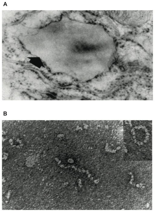
Figure 2.
(A) Electron microscopy (×20,000) of a hepatocyte from a Z homozygote showing a massive inclusion (arrowed) in the endoplasmic reticulum. Reproduced from Lomas et al18 with permission. (B) The intra-hepatic polymers of mutant Z α1-antitrypsin have the appearance of beads on a string on electron microscopy. Reproduced from Lomas et al137 with permission.

