
Figure 1. CBF during ischaemia and reperfusion and subsequent injury in the rat model of MCAO.
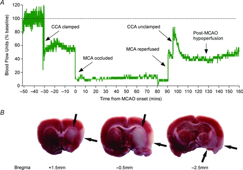
A, methodology of the MCAO technique and ethical regulations associated with these experiments have been previously described (Nagel et al. 2011). Laser Doppler flowmetry was used to measure relative CBF over the right somatosensory cortex of a male Wistar rat. Baseline CBF was normalized to 100% blood flow units (BFU). Upon temporary common carotid artery (CCA) ligation, CBF was reduced to 60% BFU. A silicon-coated 4-0 monofilament was then inserted through the external carotid artery and advanced up the internal carotid artery to occlude the right middle cerebral artery (MCA). MCAO was confirmed by a sharp decrease in CBF to < 20% BFU, and this was maintained for 90 min. Reperfusion of the MCA was achieved through retraction of the monofilament, when a sharp increase in CBF and a small hyperaemia was observed. After 5 min of MCA reperfusion (to allow removal of the monofilament), the CCA was unclamped to allow full reperfusion to the ischaemic area, which produced a further increase in CBF and hyperaemia lasting approximately 10 min. This was followed by a post-ischaemic hypoperfusion at 50% BFU during the next 60 min. B, cerebral injury was observed 24 h post-ischaemia onset in the striatum and the cortex (arrows) from the same animal as A using triphenyl-tetrazolium staining.