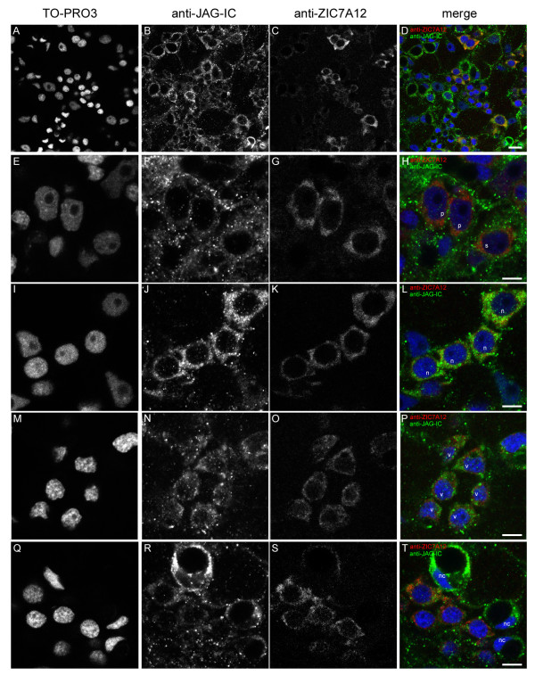
Figure 12.
Co-immunofluorescence staining with anti-JAG-IC- and anti-ZIC7A12-antibodies. Single confocal sections of a hydra from a region of the body column: (A, E, I, M, Q) DNA staining with TO-PRO3, (B, F, J, N, R) anti-JAG-IC staining, (C, G, K, O, S) anti-ZIC7A12 staining, (D, H, L, P, T) merged images of all channels in false colours: DNA (blue), anti-JAG-IC staining (green), anti-ZIC7A12 staining (red); (A-D) overview of the staining pattern in the body column of an adult hydra, (E-H) localization of HyJagged in single (s) and a pair of interstitial cells (p), (I-L) staining of a nest of four nematoblasts (n), (M-P) nest of nematoblasts with vacuoles (v), (Q-T) staining of mature nematocytes (nc); scale bars: (A-D) 10 μm, (E-T) 5 μm.

