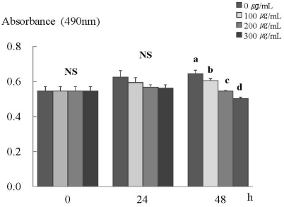
Fig. 1.
ERL inhibits cell proliferation in MDA-MB-231 cells. MDA-MB-231 cells were plated at a density of 2.5×10⁴ cells/ml in a 24 well plate with DMEM/F12 supplemented with 10% FBS. Monolayers were then serum-starved with DMEM/F12 supplemented with 5 µg/ml transferrin, 5 ng/ml selenium, and 1 mg/ml bovine serum albumin for 24 h. After serum starvation, the monolayers were incubated in serum-free medium with 0, 100, 200, or 300 µg/mL ERL for 0, 24, or 48 h. Each bar represents the mean ± S.E. of three independent experiments. Different letters indicate significant differences among groups at α = 0.05 as determined by Duncan's multiple range test.

