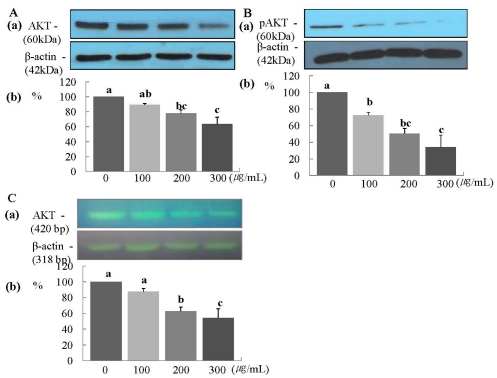
Fig. 4.
ERL reduces protein and mRNA expression of Akt MDA-MB-231 cells. For Akt and pAkt protein expression, MDA-MB-231 cells were seeded in a 100 mm dish at a density of 1×10⁵ cells/dish with DMEM/F12 supplemented with 10% FBS for 48 h. The cells were then incubated in serum free medium for 24 h, after which they were incubated in the presence of ERL at concentrations of 0, 100, 200, or 300 µg/mL for 48 h. Equal amounts of cell lysates (30 µg) were then resolved by SDS-PAGE, transferred to a membrane, and probed with Akt (A) and pAkt (B). For Akt mRNA expression, the cells were cultured in serum-free medium with ERL at concentrations of 0, 100, 200, or 300 µg/mL for 48 h. Total RNA was isolated and RT-PCR was performed (C). a) Photographs of the bands, which are representative of three independent experiments. b) Quantitative analysis of the bands. Each bar represents the mean ± SE of three independent experiments. Different letters indicate significant differences among groups at α = 0.05 as determined by Duncan's multiple range test.

