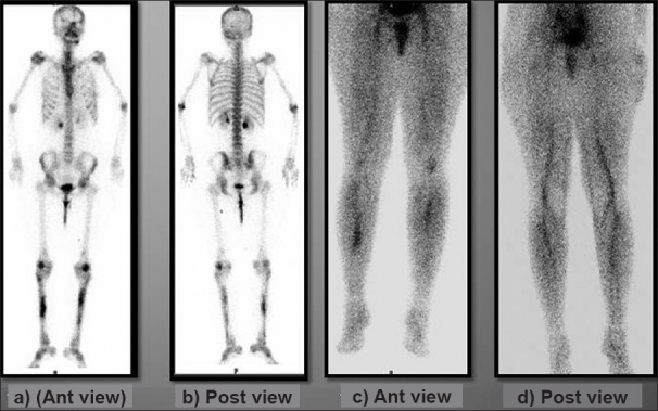
Figure 2.

(a and b) Anterior and posterior delayed phase images of 99mTc-MDP bone scan showing irregular increased uptake in the diaphyseal region of both tibiae; rest of the skeleton shows physiologic uptake. (c and d) Anterior and posterior pool phase images of 99mTc-MDP bone scan show increased uptake in the bilateral mid-calf region of both legs
