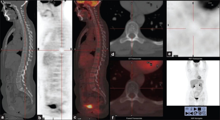
Figure 2.

Computed tomography (CT) (a), positron emission tomography (PET) (b) and PET/CT (c) sagittal slices, and CT (d) PET (e) and PET/CT (f) transversal slices. The only pathological finding at the F-18 FDG PET/CT study was an ametabolic area at the T9 vertebra on PET images. CT shows thickened vertical trabeculae on sagittal images and punctate sclerotic foci on transversal images at the body of T9. Findings are highly suggestive of vertebral hemangioma
