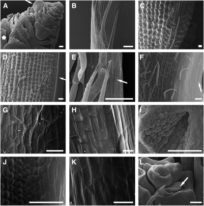
Figure 3.
Scanning Electron Microscopy Analysis of Mutant Flowers.
(A) Inner organs of a mads3-3 mads58 double mutant floral bud showing a second-whorl lodicule (asterisk), a fourth-whorl lemma-side palea-like primordium (arrow), several ectopic lodicules, and the enlarged FM.
(B) Wild-type sterile lemma with the abaxial surface oriented on the left.
(C) to (F) Abaxial surface of wild-type lemma (C), wild-type palea (D), mads3-3 mads58 double mutant palea-like organ (E), and mads3-3 mads13 mads58 triple mutant palea-like organ (F). The palea and palea-like marginal regions are shown in (D) to (F) (arrows).
(G) to (K) Adaxial surface of wild-type lemma (G), wild-type palea (H), mads3-3 mads58 double mutant palea-like organ ([I] and [J]), and mads3-3 mads13 mads58 triple mutant palea-like organ (K).
(L) Mads3-3 mads13 double mutant indeterminate FM surrounded by several developing carpel primordia and also an ectopic lodicule primordium (arrow).
Bars = 100 μm in (D) and 50 μm in the other pictures.

