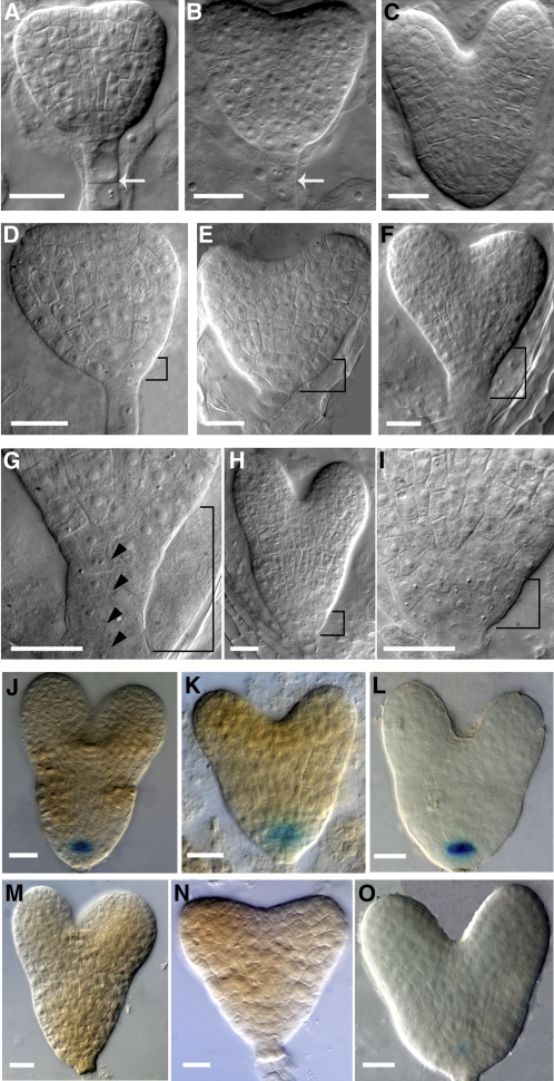
Figure 2.
Downregulation of RopGEF7 during Embryogenesis Induces Defects in Cell Division and Maintenance of the QC.
(A) to (C) Wild-type embryos in early heart (A), heart (B), and late heart (C) stages. Arrows indicate the anticlinal cell division in suspensor cells.
(D) to (I) Basal cell division defects in RPS5Apro:RopGEF7 RNAi embryos at the early heart (D), heart ([E] and [F]), and late heart (H) stages. (G) and (I) show magnifications of basal cell regions (bracketed) in (F) and (H), respectively. Arrowheads point to periclinal cell divisions in suspensor cells.
(J) to (L) GUS staining of QC25:GUS (J), QC46:GUS (K), and RopGEF7pro:GUS (L) in a control embryo.
(M) to (O) GUS staining of QC25:GUS (M), QC46:GUS (N), and RopGEF7pro:GUS (O) in an RNAi embryo.
Bars = 20 μm in (A) to (O).

