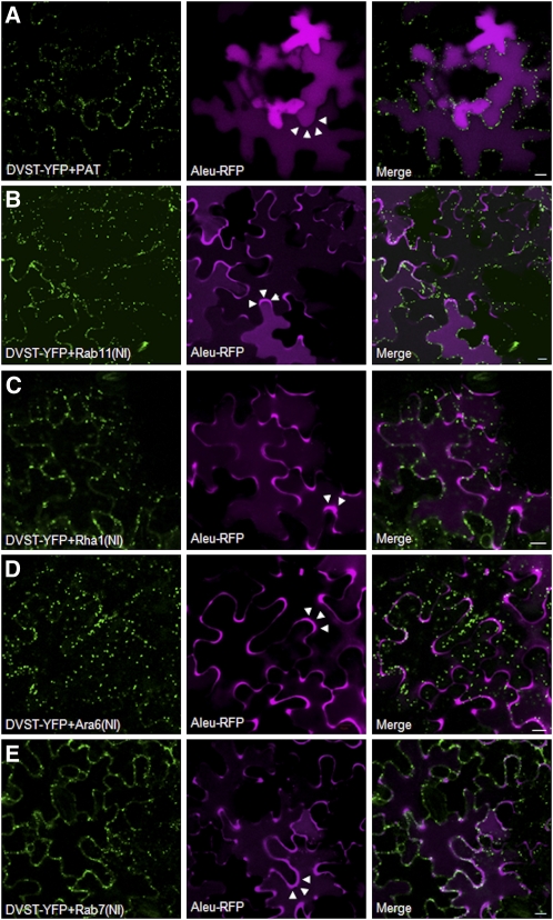
Figure 3.
The Fluorescent Chimera Aleu-RFP Is Redirected to the Apoplast in the Presence of Nucleotide-Free Variants of Rab11, Rha1, Ara6, and Rab7.
An Agrobacterium strain harboring an Aleu-RFP encoding plasmid was infiltrated together with the strain harboring the dual expression vectors encoding for the Golgi marker ST-YFP together with either PAT (A) as mock effector or Rab11(NI) (B), Rha1(NI) (C), Ara6(NI) (D), or Rab7(NI) (E) mutants as test objects.
(A) Cells expressing comparable levels of the internal standard ST-YFP were chosen for imaging the midsection of the lower epidermis to evaluate both vacuolar and apoplastic fluorescence of Aleu-RFP where appropriate. The mock effector PAT does not prevent normal vacuolar localization of Aleu-RFP, as shown by diffuse fluorescence in the central vacuole and an absence of signal in the apoplast (white arrow heads).
(B) to (E) Rab11(NI), Rha1(NI), Ara6(NI), and Rab7(NI) mutants cause mis-sorting of Aleu-RFP to the apoplast of the cells (white arrowheads). Bars = 10μm.

