Figure 8.
AMSH3 Enzymatic Activity Affects VPS2.1 and VPS24.1 Intracellular Localization.
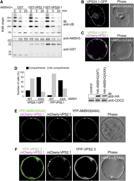
(A) AMSH3 DUB assay with purified GST, GST-VPS2.1, or GST-VPS24.1 using K48-linked ubiquitin chains. AMSH3 was preincubated with GST fusion proteins prior the addition of ubiquitin chains to the reaction mixture. Reactions were stopped after 0, 5, and 20 min and subjected to immunoblots using the indicated antibodies.
(B) and (C) Coexpression of 35Spro:HA-AMSH3(AXA) with 35Spro:VPS24.1-GFP (B) or UBQ10pro:YFP-VPS2.1 and 35Spro:VPS24.1-GFP (C). Upon expression of AMSH3(AXA), both VPS2.1 and VSP24.1 fusion proteins partially mislocalize on compartments.
(D) Quantification of the effect of 35Spro:HA-AMSH3(WT) and 35Spro:HA-AMSH3(AXA) coexpression on ESCRT-III localization (left panel). Solid bars, cells with compartments; gray bars, cells without compartments. n = 35 and 34 for 35Spro:VPS24.1-GFP expressed with 35Spro:HA-AMSH3(WT) and 35Spro:HA-AMSH3(AXA), respectively, and n = 16 and 30 for UBQ10pro:YFP-VPS2.1 expressed with 35Spro:HA-AMSH3(WT) and 35Spro:HA-AMSH3(AXA), respectively. Expression of HA-AMSH3(WT) and HA-AMSH3(AXA) was verified by immunoblotting with an anti-HA antibody (right panel). An anti-CDC2 antibody was used to show equal loading.
(E) mCherry-VPS2.1 and YFP-AMSH3(AXA) colocalized on the AMSH3(AXA)-induced compartments.
(F) mCherry-VPS2.1, but not YFP-VPS2.3, localizes on AMSH3(AXA)-induced cellular compartments.

