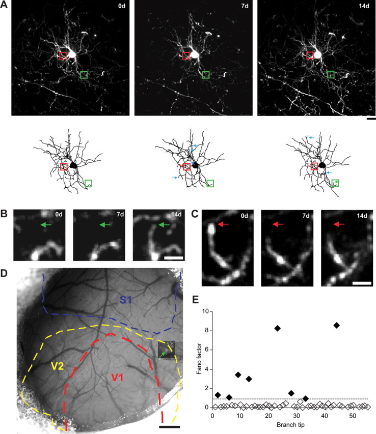
Figure 1.

Chronic two-photon in vivo imaging of dendritic branch tip dynamics in superficial L2/3 cortical interneurons. A, Maximum z-projection (MZP) near the cell body (above) along with two-dimensional projections of three-dimensional skeletal reconstructions (below) of a superficial L2/3 interneuron acquired over 2 weeks. Arrows indicate dynamic branch tips. B, High-magnification view of one branch tip extension (green box and arrow in A). Green arrow marks the approximate distal end of the branch tip at 14 d. C, High-magnification view of one branch tip retraction (red box and arrow in A). Red arrow marks the approximate distal end of the branch tip at 0 d. D, MZP of chronically imaged interneuron (green arrow) superimposed over blood vessel map with primary visual cortex (V1, red), secondary visual cortex (V2, yellow), and primary somatosensory cortex (S1, blue) outlined. E, Dynamic branch tip analysis of cell imaged in A. Diamonds indicate Fano factor values of individual monitored branch tips with filled diamonds indicating dynamic branch tips. Dotted line marks the experimentally determined threshold for dynamic branch tip. Scale bars: A, 200 μm; B, 5 μm; C, 0.5 mm.
