
Figure 3. Cellular heterogeneity and metastasis of ARCaPE orthotopic tumors after re-inoculation.
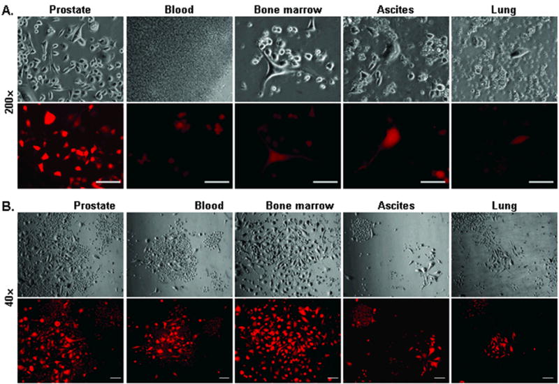
Following the first round orthotopic tumor formation, cloned ARCaPE-R31 cells were re-inoculated orthotopically into athymic mice. A, detection of metastatic tumor cells. After euthanasia due to tumor burden, samples of the prostate, blood, bone marrow, ascites, and lung were used in ex vivo culture of tumor cells. Similar results were obtained when animals bearing ARCaPE-R32 tumors were examined. All microphotographs were taken at 200x magnification, 24 hours into the culture. For each field, photos of the bright field (upper) and red fluorescence (lower) are shown. Bar = 100 μm. B, heterogeneity of the tumor cells. In this presentation, microphotographs of ARCaPE-R31 tumor cells from orthotopic tumor and metastasis sites were taken after 2 weeks of ex vivo culture. All the microphotographs are at 40x magnification to show cell size differences between colonies and between cells within the same colony. For each field, photos of the bright field (upper) and red fluorescence (lower) are shown. Bar = 100 μm.