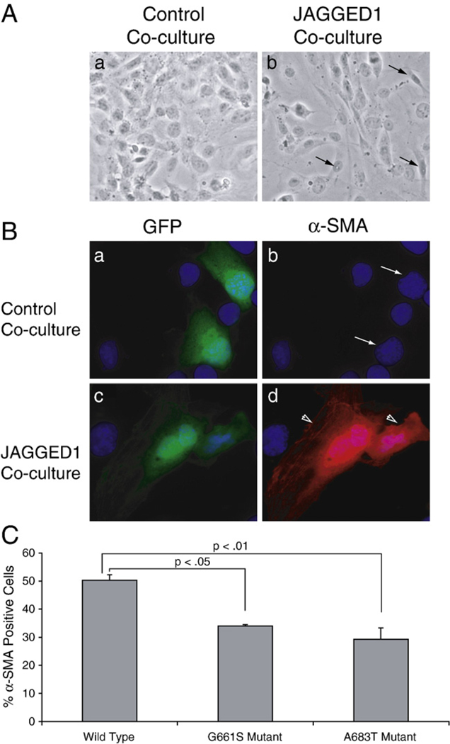
Fig. 5.
Cell lines expressing LVOT-associated NOTCH1 variants exhibit defective JAGGED1-induced EMT. A. Phase-contrast micrographs of HMECs expressing wild-type NOTCH1 co-cultured with HMEC cells containing empty vector (a) or JAGGED1 expression vector (b). When NOTCH1 expressing cells are co-cultured with JAGGED1 expressing cells, they undergo a morphological transformation characterized by loss of intercellular junctions, loss of cellular monolayer, formation of filopodia, and irregular cell shape (arrows). B. HMECs co-transfected with a NOTCH1 expression vector and a GFP expression vector were co-cultured with control HMECs (a and b) or HMECs expressing JAGGED1 (c and d). Transfected cells (as identified by GFP expression (a, c)) were examined for α-SMA staining (b and d). Cells underwent EMT only when NOTCH1 expressing HMECs were co-cultured with HMECs expressing JAGGED1 (compare arrows in b to arrowheads in d). C. The efficiency of EMT induction by JAGGED1 was compared between cells expressing wild-type NOTCH1 and cells expressing NOTCH1G661S or NOTCH1A683T. The percentage of transfected cells (as identified by GFP expression) that had also undergone EMT (as defined by morphology and α-SMA expression) was quantified. The percentages reflect the average efficiency of EMT for100 observed cells in three independent experiments. The efficiency of EMT is significantly reduced in cells that express mutant forms of the NOTCH1 receptor (p<0.05, ANOVA followed by Dunnet post hoc).

