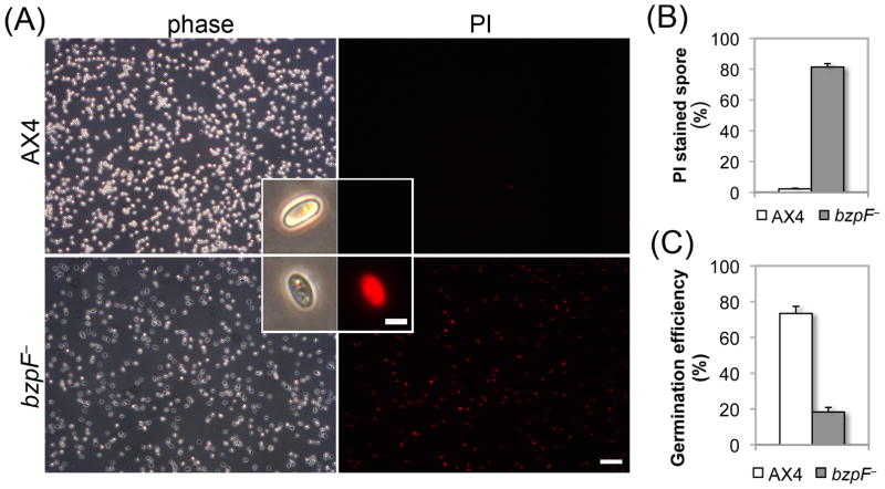
Fig. 4. bzpF– spores are compromised.
(A) PI staining of spores. Phase-contrast (phase) and fluorescence (PI) images of each field of AX4 and bzpF– spores, as indicated. Magnified spores are shown as inserts. Bars = 40 μm (low magnification) and 5 μm (inserts). (B) Quantitation of PI staining: We counted the PI-stained spores of AX4 (white bar) and bzpF– (grey bar) by flow cytometry and the fraction (%) of PI positive-spores is shown (y-axis). Data are the means + s.e.m of five replicates. (C) Germination: We incubated AX4 (white bar) and bzpF– spores in nutrient medium. The number of germinated amoebae is shown as a fraction (%) of the total spores (y-axis). The data are the means + s.e.m of three replicates.

