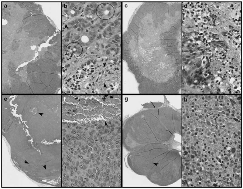
Figure 4.

Hematoxylin and eosin-stained sections of ARO (a–d) and DRO (e–h) xenograft tumors from nude mice treated with UV-inactivated (a, b, e and f) or intact ONYX-411 virus (c, d, g and h). In each case, the panels (b, d, f and h) are higher magnification images of the cells. Arrows indicate regions of blood vessel formation. The circled structures represent empty follicles with poorly organized follicular thyroid cells, which were frequently observed in ARO cell tumors (b). Similar structures were not observed in the case of DRO cell tumors (f). UV, ultraviolet.
