Figure 4.
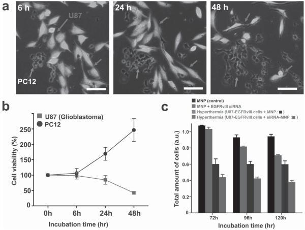
In vitro hyperthermia and siRNA-delivery studies of the FeCo/C NPs. a,b) U87–EGFP cell death induced by hyperthermia in cocultures of the highly tumorigenic U87–EGFP cells (marked by arrows) and the less-tumorigenic PC-12 cells (marked by arrows) via the targeted delivery of FeCo/C NPs to the U87 cells. Fluorescence images (a) and quantitative analysis (b) show that significant hyperthermia-induced cell death is observed in U87 cells, while the PC-12 cells keep proliferating with time. (Note that the number of cells at 0 h was taken to be 100% and the cell counts at other time points were normalized to this value.) Annexin V assays for detection of early apoptosis proved that the cell death was caused by localized hyperthermia-induced apoptosis rather than necrosis (see Figure S12 in the SI). c) MTS assay demonstrating the synergistic inhibition of proliferation and induction of cell death by the combined siRNA and hyperthermia treatment using siRNA-FeCo/C NPs in U87–EGFRvIII cells as compared to individual treatments and nontreated controls. The scale bar in all images is 100 μm.

