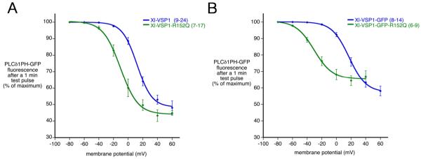
Fig. 5.
Mutation of arginine 152 to glutamine shifts Xl-VSP1 phosphatase activation to more negative membrane potentials. A: Xl-VSP1-R152Q activates at voltages ~20 mV more negative than Xl-VSP1, as measured by PLCδPH-GFP fluorescence intensity at one minute after a depolarizing test pulse. B: Xl-VSP1-GFP-R152Q activates at voltages ~50 mV more negative than Xl-VSP1-GFP, as measured by GFP fluorescence intensity at one minute after a depolarizing test pulse. The fluorescence intensity in (B) is a sum of that from PLCδ1PH-GFP and a small component from the GFP tag on the VSP (< 20%). The curves are Boltzmann fits to the averaged data. Error bars indicate mean +/− SEM, (n) = number of oocytes, listed as a range in cases where the number of data points varied for different voltages, except for Xl-VSP1-R152Q at +60 mV, where n = 3 (Fig. 5A).

